
Return to Blog
Homepage
Blood on the Brain: High Yield CT of Intracranial Hemorrhage for the USMLEr
- by
- Apr 03, 2019
- Reviewed by: Amy Rontal, MD
Identifying hemorrhage within and surrounding the brain on non-contrast CT is very high yield for all steps of the boards. Knowing a few simple features of the imaging findings for each bleed type will help you use the images to your favor and answer the associated question with ease.
One concept that applies to all acute blood on/in the brain for the sake of the boards is that blood is high density, that is it appears white on the CT images.
Epidural hemorrhage
The key feature of epidural hemorrhage is its lentiform biconvex shape. This is due to a high pressure arterial bleed which strips away the tightly adherent dura form the calvarium. Other features to notice include the inability for the bleed to cross cranial bone sutures, as well as a fracture of the temporal bone over the region of the middle meningeal artery. Look for epidural bleeds post trauma.

Case courtesy of Dr Sandeep Bhuta
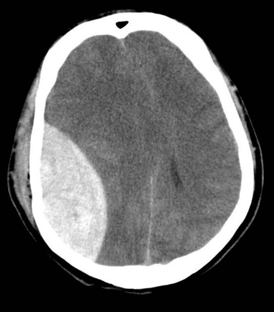
Subdural hemorrhage
The key feature of subdural hemorrhage is its crescent shape, which slides along the convexity of the cranium. Rupture of bridging veins causes this slow flow bleed. Subdural bleeds will not cross the midline at the falx due to the dural reflection of the falx keeping the blood that is accumulating underneath it from moving past the dura to the other hemisphere. Look for subdural bleeds in three populations: in the elderly, in child abuse victims, and in big trauma patients.

Case courtesy of A. Prof Frank Gaillard
Subarachnoid hemorrhage
The key feature of subarachnoid hemorrhage is that it accumulates within the sulci, ventricles, and cisterns. Essentially anywhere there is CSF, there can be subarachnoid blood. Typically on exams blood is shown in the sulci, but be aware that blood can also accumulate in the ventricles and cisterns, so if you see blood centrally in a CT it might be subarachnoid. Look for subarachnoid blood in ruptured aneurysms and in trauma.

Case courtesy of Dr. David Cuete
Intraparenchymal hemorrhage
The key feature of intraparenchymal hemorrhage is that the blood is contained within the actual brain tissue and typically has a more globular shape. Look for intraparenchymal blood in patients with hypertension. Areas of parenchymal hemorrhage can also be seen post trauma, typically in area of contusion of the anterior temporal lobes and inferior frontal lobes.

Case courtesy of Dr. David Cuete

Related Posts


Search the Blog
Try Blueprint Med School Study Planner
Create a personalized study schedule in minutes for your upcoming USMLE, COMLEX, or Shelf exam. Try it out for FREE, forever!
SIGN UPCould You Benefit from Tutoring?
Sign up for a free consultation to get matched with an expert tutor who fits your board prep needs
Get StartedFind Your Path in Medicine
A side by side comparison of specialties created by practicing physicians, for you!
Get Started