Return to Blog
Homepage
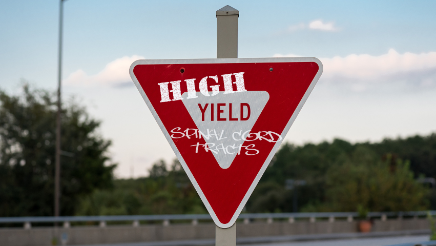
Now, That’s What I Call High-Yield: Understanding Tracts of the Spinal Cord
- by
- May 21, 2019
- Reviewed by: Amy Rontal, MD
Neurology is a huge topic on USMLE exams. There is overlap between neuro and every other body system in the grand intertwining of how we function. As such, neurology demands loads of time to study and internalize the material. One of the hallmarks of neurology is a heavy focus on anatomy. Whether we are talking about the anatomic framework of the brain, the different functions of different regions of the spinal cord, or the dermatomes and myotomes covered by different spinal nerves, anatomy is central to the understanding of neurology.
A topic which often trips students up, and one that you will be sure to be tested on, is the ascending and descending pathways that connect brain and periphery on a sensory and motor perspective. These tracts have esoteric names, non-intuitive crossing patterns, and completely different functions from one another. Keeping them compartmentalized is essential to both understanding how our nervous system is laid out, and earning more correct answers on Test Day.
Let’s delve deeper into the sensory and motor tracts of the spinal cord, and cover the information that you absolutely need to know for the USMLE.
For simplicity, we will divide these tracts into two main groups – motor and sensory. As a caveat, we are going to disregard the ~7 lesser tracts (e.g., rubrospinal, tectospinal, reticulospinal, etc.) and build a simpler model of the spinal cord. At the expense of losing the most esoteric, we are hanging onto what is most high-yield.
High-yield Motor Tracts of the Spinal Cord
The motor system is controlled by a descending tract – the corticospinal tract. Think about how we make a conscious movement. It starts with a thought – I’m going to kick this ball, or I’d like to reach for that soda. While this thought “occurs” in the prefrontal cortex, the stimulus to move a muscle is generated in the motor strip of the precentral gyrus in the frontal lobe. That’s why this is a “descending” tract. It originates in the brain, and terminates at the neuromuscular junction. Part of the magic of this tract is that all movement is coordinated by just two nerves. It’s incredible that it takes a simple two-nerve network to activate any muscle. The first-order nerve has to connect the motor strip in the brain to the cell body of the anterior horn of the spinal cord. The second-order neuron connects the anterior horn to the neuromuscular junction. From there, the transmitted action potential leads to release of acetylcholine, propagation of the action potential across the NMJ, and contraction of the muscle. I reiterate: those are the only two nerves you need to know to understand the motor system. It’s almost too good to be true.
As we progress one level deeper, we are left with tracing the anatomic pattern that these nerves take. The first order neuron has some important twists and turns on its way to the anterior horn of the spinal cord. It courses through the internal capsule in the brain, makes an important crossing to the contralateral spinal cord in the medulla of the brain stem, and courses down the lateral corticospinal tracts. This “decussation of the pyramids” is a flashy name that means motor nerves cross from right brain to left spinal cord in order to control left-sided muscles (and vice-versa). The path of the second-order motor neuron is straightforward – it exits the lateral foramen (hole) in between adjacent vertebrae, and courses to the muscle being supplied, oftentimes synapsing at a plexus (e.g., brachial, lumbar) along the way.
This tract gets its blood supply from a singular anterior spinal artery. Injury or compromise of pressure in this artery can lead to spinal cord ischemia, and if severe enough, paralysis.
High-yield Sensory Tracts of the Spinal Cord
Sensory tracts are slightly more difficult to study, as there are two important ones, and they contain three nerves in the sequence instead of just two.
Ascending (sensory) tract 1 is the dorsal columns, whose nerves are responsible for vibration, fine touch, and proprioception. While these tracts still connect body to brain, they run in the opposite direction, from periphery to brain. First-order neurons travel from periphery to the dorsal column, second-order neurons connect dorsal column to the thalamus, crossing in the medulla. High-level third-order neurons connect thalamus to the sensory strip in the postcentral gyrus of the temporal lobe.
Ascending (sensory) tract 2, the spinothalamic tract, is the wildest of them all, secondary to its kooky decussation. This tract is responsible for pain and temperature transmission. The first-order neuron, whose body lies in the DRG, connects periphery to the spinal cord, and travels one or two levels before crossing over. This decussation occurs in the anterior white commissure of the spinal cord, not in the medulla like the other tracts. Second-order neurons take the signals from the spinal cord to the thalamus, and third-order from thalamus to cortical, higher-level processing centers.
As you trace these paths in your head, think of cars traveling down highways. Take the car down the motor pathway from the brain to the to the medulla, change lanes and cross over, course down the corticospinal tract express, synapse (and honk) on the anterior horn, and cruise home to the neuromuscular junction. Close your eyes and trace out the tracts, and they will cement themselves in your mind. Knowledge of them will help explain any spinal cord lesion, and is essential to understanding different pathologies and deficits.
Want more high yield? Learn how to parse out high-yield material for your USMLE study period. Review high-yield study for the musculoskeletal system and high-yield joints review.

Related Posts


Search the Blog
Try Blueprint Med School Study Planner
Create a personalized study schedule in minutes for your upcoming USMLE, COMLEX, or Shelf exam. Try it out for FREE, forever!
SIGN UPCould You Benefit from Tutoring?
Sign up for a free consultation to get matched with an expert tutor who fits your board prep needs
Get StartedFind Your Path in Medicine
A side by side comparison of specialties created by practicing physicians, for you!
Get Started