Return to Blog
Homepage
Radiology for the USMLE: The Chest X-Ray
- by
- Jun 19, 2018
- Reviewed by: Amy Rontal, MD
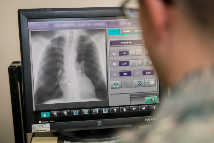
Chest X-Rays are one of the most common images given on the boards. The boards do not expect greatness when reading images: only to spot the obvious abnormality and to apply that information to the question given (see “Radiology for the USMLE: A Stepwise Approach” for a more detailed explanation of this). Here are a few of the most commonly tested findings and how they might be used as tools to answer the question:
1. Pneumonia
Pneumonias have a very large spectrum of presentations on chest radiographs. The most commonly tested presentation is the lobar pneumonia (pictured below) which looks like a wedge shaped opacity occupying the space a normal lobe of the lung would. The exact look of a pneumonia may also aid in your differential, as bugs like Streptococcus pneumoniae and Klebsiella pneumoniae tend to have a unilateral lobar appearance whereas bugs like Legionella pneumoniae and Mycoplasma pneumonia tend to be bilateral and more diffuse.
Case courtesy of Dr Roberto Schubert, Middle Lobe Pneumonia
2. Mass
Masses on chest radiographs on the boards tend to be very large and round. If there is a single mass then you should be thinking of a primary pulmonary neoplasm (pictured below), whereas if there are multiple so called “cannon-ball” like masses then you should be thinking of metastasis (pictured below).
Case courtesy of A.Prof Frank Gaillard, Lung Cancer
Case courtesy of Dr Aditya Shetty, Lung Metastases
3. Pneumothorax
The pneumothorax can be difficult to spot. The first thing to do is to take a step back and compare the two lung fields. Is one darker than the other? The darker side would be the side with the pneumothorax. Next look for a line that delineates where the lung markings stop and after there is increased darkness (see below). This line is where the pneumothorax stops and the collapsed lung begins. Most of the pneumothoraxes on the boards are very large, but even so they can be hard to see unless you take a step back. Remember, if there is significant mediastinal shift to one side in the setting of a pneumothorax then that is a tension pneumothorax and needs needle decompression ASAP.

Case courtesy of Dr Chris O’Donnell, Pneumothorax Due To Apical Blebs Surgicallly Treated
4. Pulmonary Edema
Pulmonary edema whether cardiogenic (ie CHF) or non-cardiogenic (ie ARDS) is very commonly tested on the boards. There are several key features of pulmonary edema. The most important for the boards is recognizing interstitial and alveolar edema, as well as pleural effusions. Interstitial edema can easily be thought as an increase in thickness and prominence of the interstitial markings on the chest radiograph (i.e. the white lines in the lung parenchyma) (see below). Alveolar edema is when the fluid then spills into the alevoli from the interstitium and there starts to be a fluffy white infiltrate diffusely. Lastly another feature of pulmonary edema is when the fluid then spills into the space around the lungs and presents as pleural effusions. Pleural effusions present as flat perpendicular lines on an upright chest radiograph with confluent opacity beneath the line.

Case courtesy of A.Prof Frank Gaillard, Congestive Cardiac Failure

Related Posts


Search the Blog
Try Blueprint Med School Study Planner
Create a personalized study schedule in minutes for your upcoming USMLE, COMLEX, or Shelf exam. Try it out for FREE, forever!
SIGN UPCould You Benefit from Tutoring?
Sign up for a free consultation to get matched with an expert tutor who fits your board prep needs
Get StartedFind Your Path in Medicine
A side by side comparison of specialties created by practicing physicians, for you!
Get Started