Return to Blog
Homepage
USMLE Step 1 & Step 2 CK Question Breakdown: 2-in-1 Edition
- by
- Sep 12, 2018
- Reviewed by: Amy Rontal, MD
As a treat today, we have a question that is quite pertinent for BOTH Step 1 and Step 2 CK. A question like this could certainly appear on either test. Take a stab at it and get your learn on.
An 83-year old woman presents to the ED with very acute abdominal pain for 1 hour. She recalls that it started almost exactly at 8:45 PM. She has had similar pain in the past, but never this intense. She has a past medical history of coronary artery disease with myocardial infarction 6 years ago, hyperlipidemia, hypertension, diabetes, and atrial fibrillation. She has never had surgery before. She denies any hematemesis or dark colored stools. Her home medications include metoprolol, warfarin, simvastatin, and hydrochlorothiazide, as well as a few herbal supplements she cannot recall. She is somewhat noncompliant with her medications. The pain is an 8/10 in severity, and is a dull abdominal ache without radiation. Vital signs are HR 110, BP 147/92, T 99.0, RR 22, SpO2 98% ORA. Physical exam is significant for a diffuse abdominal pain with palpation. Previous evaluation of her pain during an ED visit 5 months prior showed no evidence of cholelithiasis.
What test would be most likely to establish her diagnosis?
- Chest X-Ray
- Abdominal CT Angiography
- RUQ US
- Amylase, Lipase, and LFTs
- MRCP
- Abdominal X-Ray
Like any question, we will start with the one-liner and form a broad differential diagnosis from there. Let us never forget how much important information is contained in this first sentence!
We’ve got:
An elderly woman (Very different set of medical problems than a 35 year old),
Presenting to the ED (Very different than a routine office visit),
With Acute Abdominal Pain (Has its own particular differential).
What comes to mind for acute abdominal pain, severe enough to warrant an ED visit, in an older female patient? Let’s go around the horn of abdominal organs. Pancreatitis? Perhaps. Gastritis/peptic ulcer disease? Definite possibility, though less likely with no blood in the GI tract. Acute hepatitis? Unlikely. Small bowel obstruction? Good chance, especially if she’s had previous abdominal surgery, but she hasn’t! Perforated viscus? Also a possibility. Ovarian pathology, like a cyst? It’s on there. Appendicitis? Not the classic age, but we’ll keep it in mind. Gallbladder pathology? Definitely could be, but her stone-free history makes it less likely. Let us not forget the possibility of a myocardial infarction presenting as abdominal pain; these non-classical, non-chest pain presentations are more common in women and diabetics. That’s relatively exhaustive. Abdominal pain is a tricky one just because the differential can be so broad. We will depend on the further information to start narrowing us down.
This poor patient has a laundry list of common medical problems. She has hypertension, hyperlipidemia, diabetes, coronary artery disease, and a previous MI. All of these conditions are likely wreaking havoc on her vasculature. She clearly has atherosclerotic disease, as evidenced by her CAD, and multiple risk factors for depositing plaque in blood vessels. Diabetes is infamous for causing micro- and macro-vascular damage, hypertension beats up the intima with every beat of the heart, and hyperlipidemia provides the substrate for laying down tracks of plaques. Remember that plaque in coronaries often means plaques in other important arteries throughout the body, including the splanchnic vasculature, carotids, femorals, and distal arteries of the legs (did someone say claudication?).
We see she has a virgin abdomen, making SBO secondary to adhesions less likely. As you recall from your surgery clerkships, adhesions top the ranks of causes of SBO, followed by hernias and malignancies.
Moving on, let’s review this meds list. She’s taking the usual cardiac patient cocktail of beta-blocker (metoprolol), antihypertensive (hydrochlorothiazide), statin (simvastatin), and in her case, likely in the setting of the thrombogenic atrial fibrillation, she is also on warfarin. The herbal supplements are a real wildcard, and depending on what they are, can interfere with liver cytochromes, making medications more or less effective, or exhibit their own effects. We also are given a history of noncompliance, a particularly unfortunate thing for a patient who is beta-blocked and anticoagulated.
Lastly, her physical exam: severe, dull, non-radiating pain. She’s tachycardic and hypertensive, a combination usually seen in patients suffering from pain. While she’s tachypneic, saturations are fine. The abdominal exam is relatively non-specific. If anything, she doesn’t have any focal tenderness in any quadrant or region.
Now, as (almost) always, we must make a diagnosis, and figure out which test will rule in or out said diagnosis. Vasculopathic, elderly lady, has a beat-up heart, non-compliant with anticoagulation (ergo, pro-thrombotic), with acute abdominal pain after having similar chronic bouts of abdominal pain. Have you pieced it together? If not, let’s go through the answer choices one-by-one and reverse engineer what diagnoses might be served by each study. It’s generally better to establish your own diagnosis, but as a backup plan, answer choices can serve as a starting point.
Chest X-Ray
While we don’t think anything pulmonary is going on, a Chest X-Ray might reveal air under the diaphragm. This could be a sign of a perforated viscus, but we don’t really have a great story for a hole in an organ. There’s no history of dilation, bleeding/ulceration, or obstruction. Probably not super useful.
Abdominal CT Angiography
Vasculature imaging of the abdomen? This would give us a shot of the aorta, renal arteries, splanchnic arteries (celiac, SMA, IMA), and iliac-femoral system. We know she has a history of atherosclerotic vessels, and atop that, is at risk for clot formation. Let’s keep this one in mind.
RUQ US
The classic imaging test for gallbladder pathology. The patient doesn’t have any specific RUQ pain, no history of biliary colic, and no stones in a recent evaluation. While the pain pattern could match acute cholecystitis atop previous biliary colic, we would probably expect some complaint from the RUQ.
Amylase, Lipase, and LFTs
Could we be looking at acute on chronic pancreatitis? Perhaps, but we really don’t have any history pointing us in that direction. Without history of gallstones, alcohol intake, new drugs, or scorpion bites, her pancreas is likely AOK. Could an herbal supplement be causing it? Low likelihood, but always something to keep in mind.
MRCP
Evaluating her biliary tree with an MRCP would be useful if we suspected some sort of pathology like cholecystitis, choledocholithiasis, or some autoimmune inflammation of the bile duct, but we have no reason to believe such. The patient’s risk factors are more likely to cause other damage.
Abdominal X-Ray
These are great for detecting dilation or obstruction, neither of which we are expecting here. They should not be ordered for vague abdominal pain.
After surveying the choices, the most likely problem with this patient is acute mesenteric ischemia. Her poor anticoagulation and atrial fibrillation likely caused generation of a thrombus in the left atrium that embolized to an already narrowed, atherosclerotic lumen of the splanchnic arteries. This explains the hyperacute nature of the abdominal pain. Now, if the stem included “pain out of proportion to exam,” it would be much too simple. Keep this relatively rarer disease etiology in mind when confronted with an older vasculopathic patient. Arterial embolism generally causes severe pain very quickly, as we see in her case.
Keep those differentials broad, and work systematically. It is then that the answer will find you.

Related Posts


Search the Blog
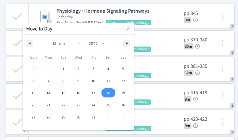
Try Blueprint Med School Study Planner
Create a personalized study schedule in minutes for your upcoming USMLE, COMLEX, or Shelf exam. Try it out for FREE, forever!
SIGN UPCould You Benefit from Tutoring?
Sign up for a free consultation to get matched with an expert tutor who fits your board prep needs
Get StartedFind Your Path in Medicine
A side by side comparison of specialties created by practicing physicians, for you!
Get Started